Home

spill Ganske London blood smear labeled Melkehvit klokke Klar

Blood film - Wikipedia

PDF] Microscopic Blood Smear Segmentation and Classification Using Deep  Contour Aware CNN and Extreme Machine Learning | Semantic Scholar
PDF] Microscopic Blood Smear Segmentation and Classification Using Deep Contour Aware CNN and Extreme Machine Learning | Semantic Scholar
Blue Histology - Blood

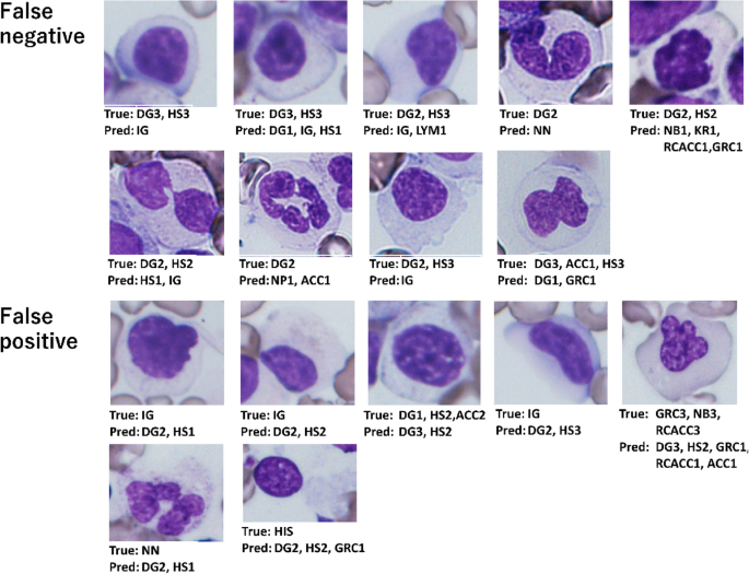
Assessment of dysplasia in bone marrow smear with convolutional neural  network | Scientific Reports
Assessment of dysplasia in bone marrow smear with convolutional neural network | Scientific Reports

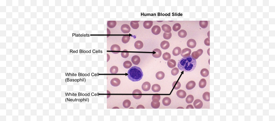
Download Top Images For Blood Smear - Labeled Mammalian Blood Smear Png,Blood  Smear Png - free transparent png images - pngaaa.com
Download Top Images For Blood Smear - Labeled Mammalian Blood Smear Png,Blood Smear Png - free transparent png images - pngaaa.com

Red Blood Cell Count- Understand the Test

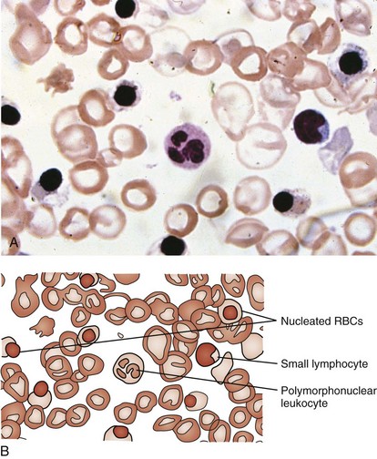
Photomicrograph of a Human Blood Smear, Wright's stain (610x) Diagram |  Quizlet
Photomicrograph of a Human Blood Smear, Wright's stain (610x) Diagram | Quizlet

Accurate Machine-Learning-Based classification of Leukemia from Blood Smear  Images - Clinical Lymphoma, Myeloma and Leukemia
Accurate Machine-Learning-Based classification of Leukemia from Blood Smear Images - Clinical Lymphoma, Myeloma and Leukemia

Types of blood cells | Kenhub

Digitalization microscopic peripheral blood smear | Download Scientific  Diagram
Digitalization microscopic peripheral blood smear | Download Scientific Diagram

Peripheral blood smear analysis using image processing approach for  diagnostic purposes: A review - ScienceDirect
Peripheral blood smear analysis using image processing approach for diagnostic purposes: A review - ScienceDirect

A dataset of microscopic peripheral blood cell images for development of  automatic recognition systems - ScienceDirect
A dataset of microscopic peripheral blood cell images for development of automatic recognition systems - ScienceDirect

Draw the structure of blood or microscopic structure / smear seen of human  blood and label its four component.
Draw the structure of blood or microscopic structure / smear seen of human blood and label its four component.

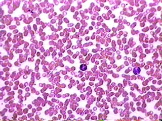
normal blood smear | Medical Laboratories

Solved This micrograph shows a peripheral blood smear | Chegg.com

Classic image: peripheral blood smear in a case of Plasmodium falciparum  cerebral malaria | BMJ Case Reports
Classic image: peripheral blood smear in a case of Plasmodium falciparum cerebral malaria | BMJ Case Reports

Blood smear, human
Blood smear, human

Hematology Society HS - Blood cells in Peripheral Blood Smear | Facebook

Hematology and Oncology | Obgyn Key

Blood Smear Preparation | Clinician's Brief

Blood smear from sea bass. Magnification 400X. | Download Scientific Diagram

Peripheral blood smear analysis using image processing approach for  diagnostic purposes: A review - ScienceDirect
Peripheral blood smear analysis using image processing approach for diagnostic purposes: A review - ScienceDirect
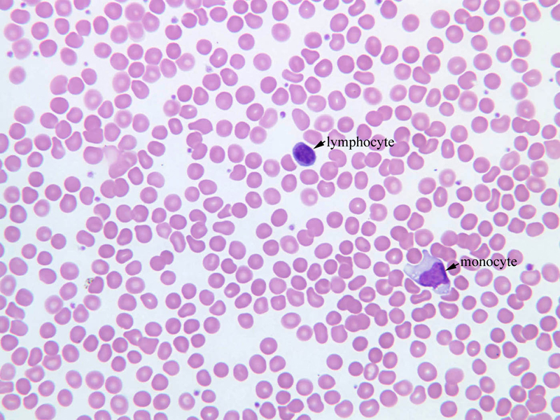
Histology - Human Blood Smear Labeled Transparent PNG - 1200x900 - Free  Download on NicePNG
Histology - Human Blood Smear Labeled Transparent PNG - 1200x900 - Free Download on NicePNG

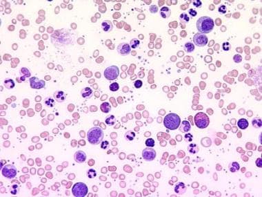
Chronic Myelogenous Leukemia (CML) Workup: Approach Considerations, Blood  Count and Peripheral Smear, Bone Marrow Analysis
Chronic Myelogenous Leukemia (CML) Workup: Approach Considerations, Blood Count and Peripheral Smear, Bone Marrow Analysis

Smear Quotes. QuotesGram

CONNECTIVE TISSUE - BLOOD BLOODFORMING TISSUES

Label-free hematology analysis using deep-ultraviolet microscopy | PNAS